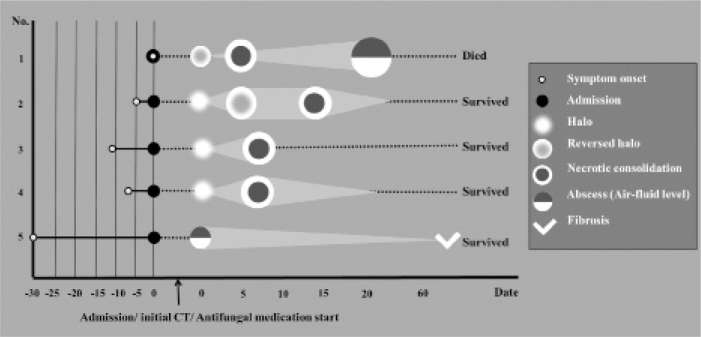
Figure 1.

The brief clinical course and morphological changes during follow-up in five patients. All patients underwent initial CT on the day of admission and started antifungal medication. Although not all cases demonstrated the same morphological features simultaneously, most tended to show consistent sequential morphological changes over follow-ups. Note that the light gray triangle or quadrangle behind the round figure represents the size change of the lesions over follow-ups. Three patients (cases 2, 4, and 5) survived and their lesions were partially resolved on follow-up chest radiographs. The patient of case 3 was transferred to an outside hospital with clinical improvement. However, the follow-up chest radiographs of this patient could not be evaluated.
