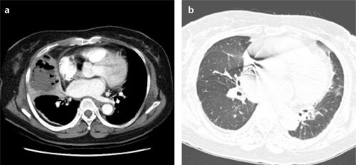
Figure 4. a, b.

(Case 5) Pulmonary mucormycosis showing abscess formation with air-fluid level in a 73-year-old female patient with diabetes mellitus. Initial chest CT (a) shows dense consolidation with internal air-fluid level in the right middle lobe of the lung, representing the internal necrosis and abscess formation. Follow-up CT taken two years later (b) demonstrates no substantial residual lesions except for mild fibrosis in the right middle lobe.
