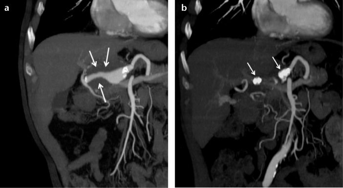
Figure 1. a, b.

Coronal CT angiography reconstruction images of a 63-year-old male show a partially thrombosed common hepatic artery aneurysm (a, arrows) and two AVPs placed in the proximal and distal parts of the aneurysm (b, arrows) one month after the occlusion.
