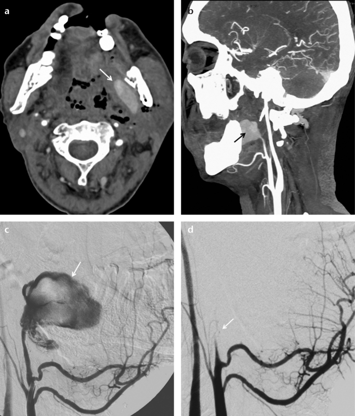
Figure 2. a–d.
Axial CT angiography image (a) of a 47-year-old male shows a pseudoaneurysm (arrow) in the left parapharyngeal space. A sagittal CT angiography reconstruction image (b) shows a left internal maxillary artery pseudoaneurysm (arrow). Selective left external carotid angiography (c) shows active contrast extravasation from the pseudoaneurysm (arrow). Selective left external carotid angiography after the deployment of an 8 mm AVP 2 (d) shows total embolization of the left internal maxillary artery (arrow).

