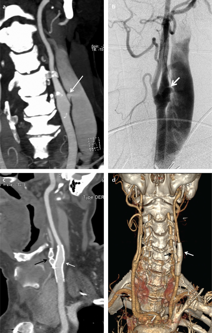
Figure 3. a–d.
Coronal CT angiography reconstruction image (a) of a 62-year-old male shows a caroticojugular fistula (arrow) between the internal carotid artery and internal jugular vein. Carotid angiography (b) shows the caroticojugular fistula (arrow). One month following the embolization, a sagittal CT angiography reconstruction image (c) shows the occluded left external carotid artery with an 8 mm AVP 4 (black arrow) and the occluded fistula with a stent-graft (white arrow). One month following the embolization, a coronal CT angiography reconstruction image (d) shows the occluded left external carotid artery with the AVP 4 (black arrow) and the occluded fistula with the stent-graft (white arrow).

