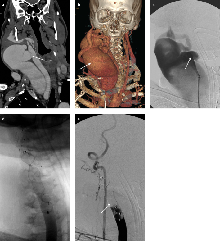
Figure 4. a–e.
Coronal CT angiography reconstruction image (a) of a 57-year-old male shows a caroticojugular fistula (arrow) between the common carotid artery and internal jugular vein. A coronal CT angiography reconstruction image (b) shows the dilated internal jugular vein (arrow) secondary to the caroticojugular fistula. Carotid angiography (c) shows the caroticojugular fistula (arrow). Carotid angiography after deployment of the AVPs (d) shows multiple AVPs. Carotid angiography after deployment of the AVPs (e) shows total embolization of the common carotid artery (arrow).

