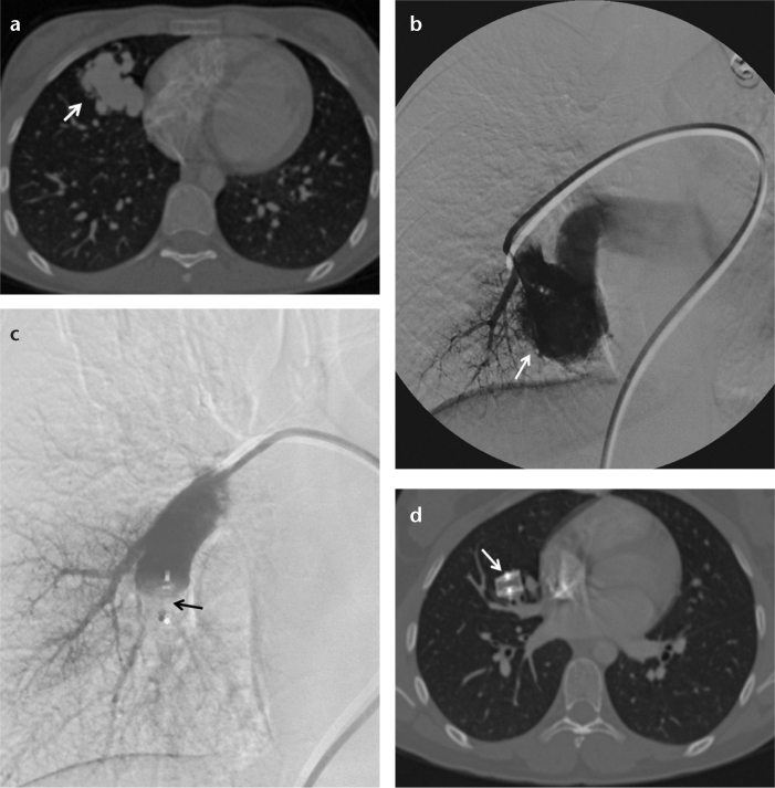
Figure 5. a–d.
Axial CT angiography (a) and pulmonary angiography (b) images of a 17-year-old female show a pulmonary arteriovenous fistula (arrow) at the right middle lobe. Pulmonary angiography after deployment of a 16 mm AVP 2 (c) shows total occlusion of the feeding artery and the AVP 2 (arrow) in the right middle lobe segment pulmonary artery. Six months later, an axial CT angiography image (d) shows the AVP 2 (arrow) in the right middle lobe segmental pulmonary artery.

