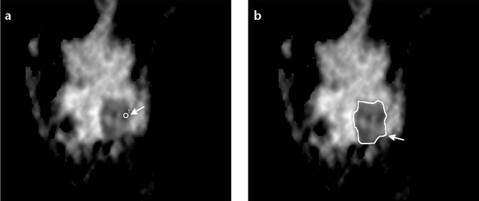
Figure 1. a, b.

A 32-year-old woman with a suspected mass lesion of 28 mm in the lower outer quadrant of the left breast. Small ROI superimposed with the ADC map in the area with the most restricted diffusion (a, white arrow). Large ROI ADC including the whole lesion (b, white arrow). Histological result was malignant lesion not otherwise specified.
