Figure 2.

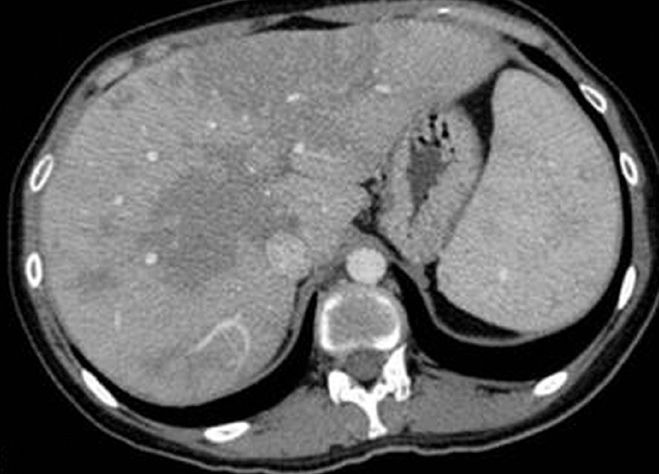
Axial contrast-enhanced CT image of a 23-year-old female with biopsy-proven hepatosplenic sarcoidosis shows coalescence of granulomas in the form of large, geographic, hypoechoic, hypodense areas in an enlarged liver and the spleen.

Axial contrast-enhanced CT image of a 23-year-old female with biopsy-proven hepatosplenic sarcoidosis shows coalescence of granulomas in the form of large, geographic, hypoechoic, hypodense areas in an enlarged liver and the spleen.