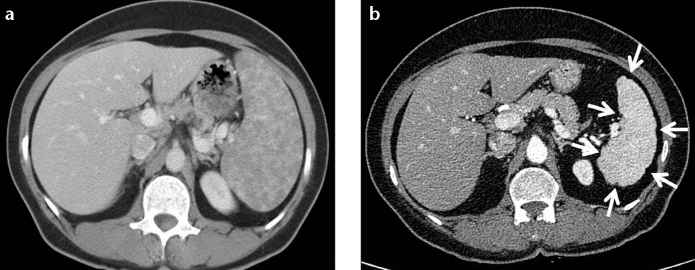
Figure 7. a, b.

A 45-year-old female with proven splenic sarcoidosis. Contrast-enhanced CT of the abdomen shows multiple hypodense granulomas infiltrating the enlarged spleen (a). Contour irregularity of the shrinking spleen became prominent within six-years follow-up (b, arrows).
