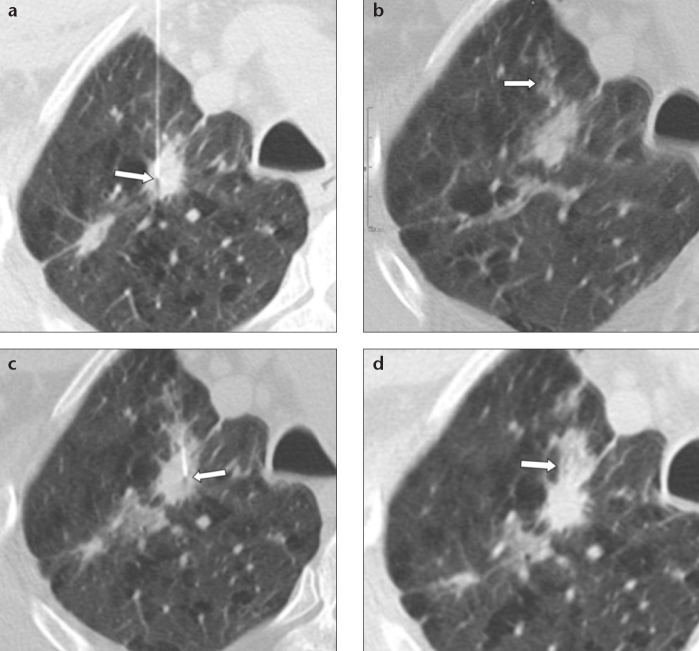
Figure 5. a–d.
CT images of an 86-year-old male smoker with solid noncalcified pulmonary nodule in the right upper lung lobe, in the presence of diffuse centrilobular and paraseptal emphysema. Panel (a) shows TTFNA of the pulmonary nodule (arrow). Panel (b) shows pulmonary hemorrhage along the needle track in the absence of pneumothorax (arrow). In the absence of a cytological diagnosis “on site”, a second sampling is carried out with reasonable safety in a different area of the pulmonary nodule (c, arrow). Note how pulmonary hemorrhage along the needle tract is increased after the second cytology (d, arrow) and pneumothorax is not present. Cytological result of TTFNA was pulmonary adenocarcinoma.

