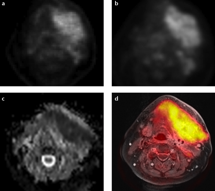
Figure 3. a–d.
Images of a large cancer of unknown primary of the head and neck region derived from PET/MRI. This example demonstrates visually comparable quality of PET/CT (a) and PET/MRI (b) derived PET images. Simultaneous PET/MRI offers the possibility of morphological, functional and metabolic imaging studies in a single examination (c, d). Diffusion-weighted imaging (ADC map; b=[0, 500, 1000]) of the lesion is shown in (c). Fusion of T1-weighted contrast-enhanced image with fat saturation and attenuation-corrected FDG-PET is shown in (d).

