
Figure 6. a–c.
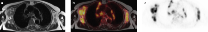
Simultaneous FDG-PET/MRI in a 72-year-old patient with an initial diagnosis of highly malignant B-cell type non-Hodgkin lymphoma. The anatomical dataset shows multiple enlarged axillary, mediastinal, as well as hilar lymph nodes (a, T2-weighted HASTE) with elevated FDG-uptake in PET (b, fusion; c, FDG-PET).