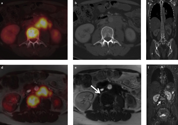
Figure 7. a–f.
A patient with bone metastases originating from ovarian cancer and local bone marrow infiltration in the lumbar spine. While in FDG-PET/CT (a–c) the bone marrow infiltration is only detectable in PET (a) and shows no osseous destruction in CT (b, c), in FDG-PET/MRI (d–f) the infiltration is detectable both in PET (d) and in MRI due to a low signal in T1-weighted sequences (e, arrow) and high signal in T2-weighted sequences (f, arrow). Especially in patients undergoing chemotherapy bone metastases/bone marrow infiltrates might not be FDG-avid and therefore not detectable in PET/CT. In this case PET/MRI might represent an advantage in whole-body imaging.

