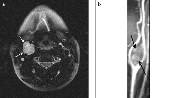
Figure 5. a, b.

Fat-saturated T2-weighted axial MRI (a) of a 36-year-old female patient shows a hyperintense glomus caroticum (arrows), typically located in the carotid bifurcation. Postcontrast coronal MR angiography arterial phase image (b) shows that the hyperintense lesion splays the internal and external carotid arteries symmetrically.
