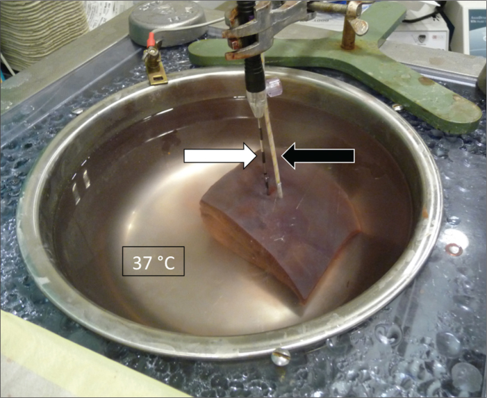
Figure 4.
RFA setup: metal bowl filled with 0.9% saline, heated to 37°C to simulate physiological body temperature, current flow and heat conduction, cuboid liver specimen, and RF electrode (white arrow). To further validate the temperature inside the parenchyma, a fiberoptic measuring system (Neoptix, Québec, Canada; black arrow) was positioned inside the parenchyma 1 cm from the expected center of each lesion.

