
Figure 2. a–c.
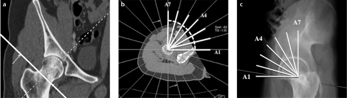
Coronal reformatted CT (a) image showing the reference plane (solid line) for radial alpha angle (AR) reconstruction. Axial CT image (b) used for the formation of radial reformatted images demonstrates superimposed radial reference lines at 15° intervals. Sagittal thick slab multiplanar reformation image (c) shows planes of radial reformats passing through the anterior, anterosuperior, and superior portions of the femoral head-neck junction.