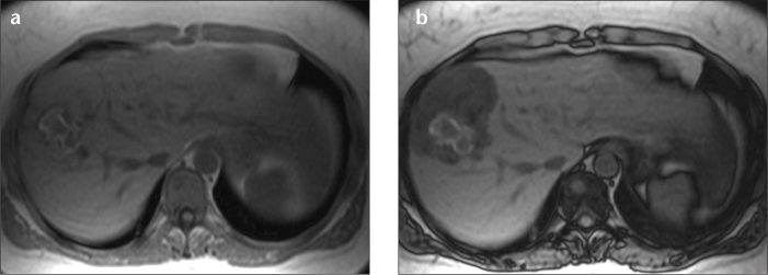
Figure 3. a, b.

Axial T1-weighted in-phase (a) and opposed-phase (b) MR images of a hepatocyte nuclear factor 1α (HNF1A)-mutated HCA. This lesion shows the typical diffuse and homogeneous suppression of signal in the lesion due to fat accumulation. Also note the blood residue centrally located in the lesion as a T1-weighted hyperintense zone. In addition to steatosis, this lesion showed no liver fatty acid binding protein staining upon histology, which is a very sensitive marker for HNF1A mutation.
