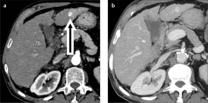
Figure 1. a, b.

A 67-year-old male with hepatitis C-related cirrhosis and capillary hemangioma. Axial contrast-enhanced CT images show a lesion (a, arrow) in the left hepatic lobe with flash-filling enhancement in the hepatic arterial phase, and isoattenuation to blood vessels in both arterial (a) and hepatic venous (b) phases.
