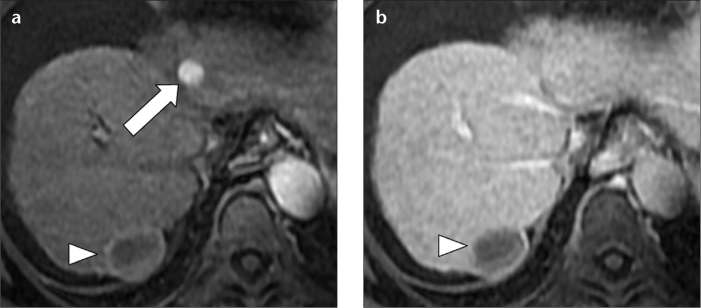
Figure 2. a, b.

Gadolinium ethoxybenzyl diethylenetriamine pentaacetic acid (Gd-EOB-DTPA)-enhanced MR images in a 71-year-old female with hepatitis C-related cirrhosis and small atypical HCC. Hepatic arterial phase fat saturated volumetric T1-weighted image (a) shows a small, hypervascular nodule (arrow) in the left hepatic lobe. Hepatic delayed phase fat-saturated volumetric T1-weighted image (b) at the same level shows isointensity of the lesion to the liver parenchyma. The lack of washout did not allow a confident diagnosis of HCC. In this patient, the diagnosis of HCC was based on lesion hypointensity in the hepatobiliary phase image (not shown). Note the hypointense lesion (arrowheads) in segment VII, representing complete necrosis of the hepatocellular carcinoma after transcatheter arterial chemoembolization.
