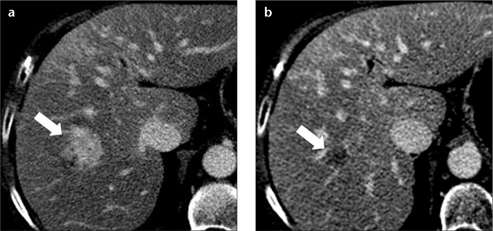
Figure 3. a, b.

A 70-year-old female with alcoholic cirrhosis and shrinking hemangioma. Hepatic venous phase CT image (a) shows a lesion (arrow) in the right hepatic lobe, which demonstrated nearly complete homogeneous enhancement with isoattenuation to the intrahepatic blood vessels. Hepatic venous phase image (b) through the same level obtained three years later, demonstrating a substantial decrease in the size of the lesion, which showed a lack of contrast enhancement. Note the absence of characteristic CT features of hemangioma in the later CT scan.
