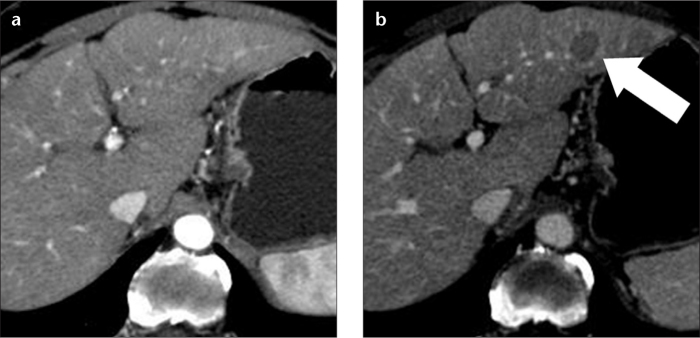
Figure 4. a, b.

A 73-year-old female with hepatitis C-related cirrhosis and hypovascular HCC in the left hepatic lobe. A hepatic arterial phase CT image (a) does not show any lesion. On a hepatic venous phase CT image (b), the lesion (arrow) is spherical and hypoattenuating to the liver parenchyma. This lesion was new in comparison with an MR examination performed one year earlier and was interpreted as hypovascular HCC, although these imaging features are also compatible with a high-grade dysplastic nodule.
