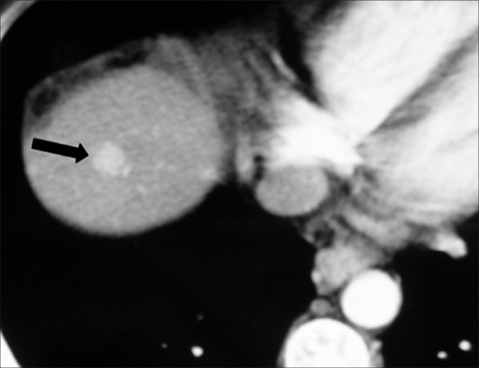
Figure 5.

A 38-year-old male with hepatitis C-related cirrhosis and focal nodular hyperplasia. Hepatic arterial phase CT image shows a homogeneously enhancing lesion (arrow) in the right hepatic lobe. This lesion was prospectively misinterpreted as HCC. The patient was transplanted, and the diagnosis of focal nodular hyperplasia was made only at explantation.
