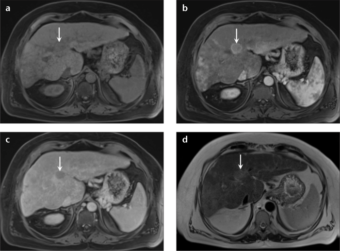
Figure 1. a–d.
A 50-year-old man with hepatitis C infection and HCC, and prior negative screening US. Axial precontrast (a), arterial (b), and delayed phase (c) T1-weighted 3D GRE images, and T2-weighted single-shot image without fat suppression (d) demonstrate an isointense lesion in segment VIII on precontrast T1-weighted 3D GRE (a, arrow), which shows avid enhancement on the arterial phase (b, arrow) and washout on the delayed phase (c, arrow) with an enhancing capsule. Mildly increased signal is observed on T2-weighted nonfat-saturated single-shot images (d, arrow); all these features are characteristic of HCC. The tumor burden was within the Milan criteria, and the patient underwent a successful transplantation.

