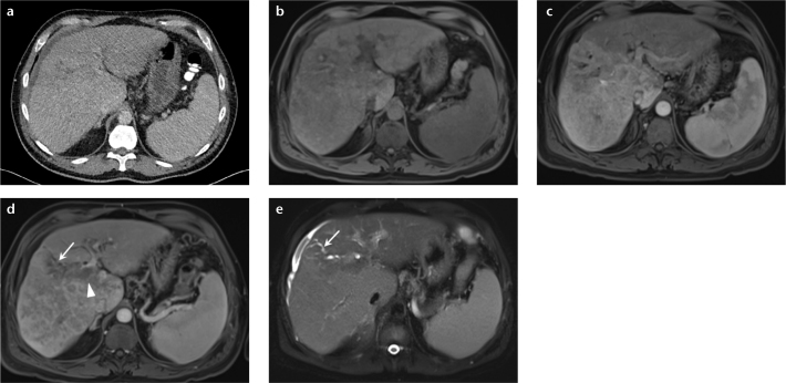
Figure 2. a–e.
A 50-year-old man with abdominal pain. Axial single phase CT from another institution (a) shows slight irregularity of the hepatic surface, mild biliary dilatation, and trace ascites. MRI of abdomen was subsequently performed to identify the cause of biliary obstruction. Unenhanced T1-weighted 3D GRE image (b) demonstrates heterogeneous signal pattern to the hepatic parenchyma. Gadolinium-enhanced arterial phase image (c) shows diffuse, nonuniform enhancement throughout the right hepatic lobe, but without a focal lesion. Delayed-phase postcontrast image (d) shows heterogeneous, nonuniform pattern of enhancement and areas of washout. Single-shot, fat-suppressed axial T2-weighted image (e) reveals abnormal, geographic regions of elevated T2 signal representing diffuse infiltrating HCC. Tumor thrombus is observed to extend into the main portal vein (d, arrowhead). Infiltrative tumor obstructs the biliary duct in the right lobe of the liver causing segmental biliary ductal obstruction (d, e, arrows).

