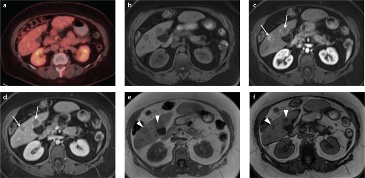
Figure 7. a–f.
A 61-year-old woman with hepatic lesions, cancer workup. Multiple biopsies returned a diagnosis of neuroendocrine metastases. PET-CT (a) showed no increased fluorodeoxyglucose avidity in the hepatic lesions. Precontrast (b) and arterial phase (c) T1-weighted 3D GRE images show two arterially enhancing masses in the right lobe of the liver, which show prompt washout on delayed phase (d). Dual echo T1-weighted GRE images (e, f) show focal areas of signal loss on the out-of-phase images (e, arrowheads) compared with the in-phase images (f, arrowheads), depicting internal lipid within the tumor. These findings are most consistent with HCC, despite the biopsy results. Special stains for HCC confirmed the diagnosis in the surgical specimen. This case demonstrates the value of MRI when used as a primary diagnostic modality for liver lesions, and HCC in particular. MRI may obviate the need for additional imaging tests and biopsy, potentially reducing overall cost while retaining high diagnostic accuracy.

