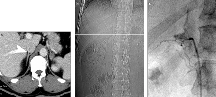
Figure 1.
a–c. Example of an adrenal vein mapping. Axial contrast-enhanced CT scan (a) gives excellent visualization of the right adrenal vein (RAV, arrowhead) located in Level 10. CT scout image (b) displays the slice position (white line) corresponding to (a). The angiogram (c) shows excellent correlation between the location of the RAV during adrenal vein sampling and the position that was found in adrenal vein mapping.

