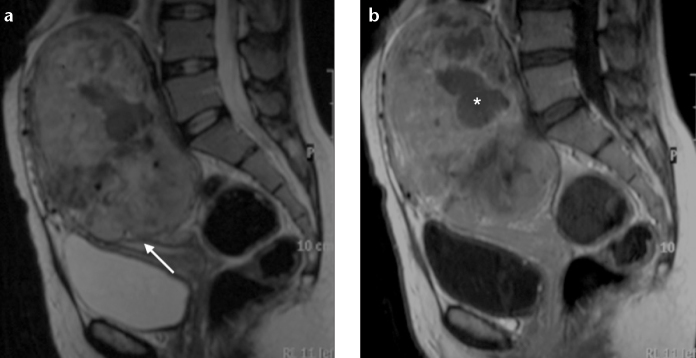
Figure 5.

a, b. Undifferentiated endometrial sarcoma in a 36-year-old woman. Sagittal T2-weighted image (a) and T1-weighted image after gadolinium administration (b) show marked uterine enlargement due to a large polypoid heterogeneous tumor, with some nodular marginality (arrow). The lesion shows intense and heterogeneous contrast uptake (uncommon for endometrial carcinoma), with a hypointense area (asterisk) suggestive of necrosis.
