FIGURE 10.

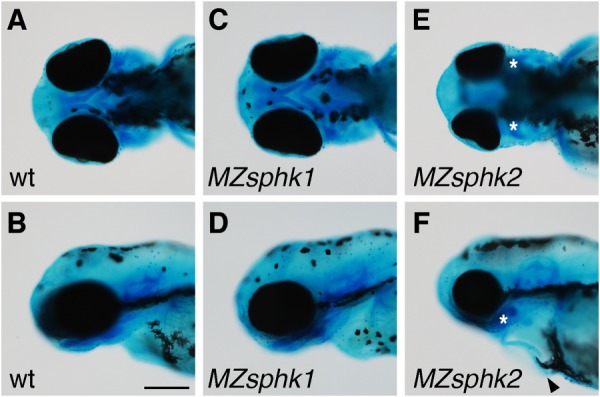
Lower jaw morphology. A–F, lower jaw morphology of wt (A and B), MZsphk1 (C and D), and MZsphk2 embryos (E and F) at 3 dpf was visualized with Alcian blue staining (A, C, and E, ventral view; B, D, and F, lateral view). The ventral pharyngeal arches of MZsphk2, but not MZsphk1 embryos, were disorganized; vestiges are shown as asterisks. Cardiac edema was also observed in MZsphk2 embryos, as shown by the black arrowhead (F). Scale bar, 200 μm.
