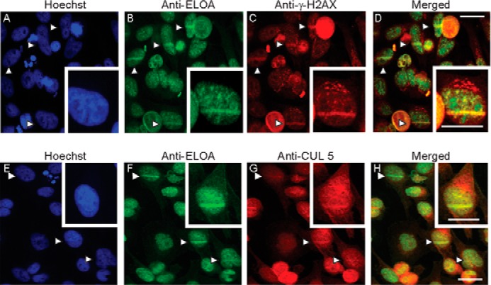
FIGURE 2.

Enrichment of Elongin A and CUL5 at sites of localized DNA damage induced by laser microirradiation. HeLa cells were stained with Hoechst dye, and some cells in each field were microirradiated with a 405-nm UV laser. Cells were fixed and analyzed by indirect immunofluorescence using anti-Elongin A (B and F) and anti-γ-H2A.X (C) or anti-CUL5 (G). Merged images are shown in D and H, and Hoechst staining is shown in A and E. Arrows, microirradiated regions. Scale bars, 26 μm.
