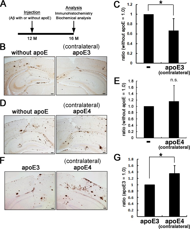
FIGURE 2.
In vivo effects of apoE on the seeding effects of Aβ protofibrils. A, schematic representation of the timeline of experiments. A7 mice were injected with Aβ protofibrils preincubated with or without apoE into the neocortex and hippocampus at 12 months. At 4 months after injection, both hemispheres were immunohistochemically or biochemically analyzed. B and C, A7 mice were injected with Aβ protofibrils preincubated without apoE on one side of the neocortex and hippocampus and those with apoE3 on the contralateral side. The both hemispheres were immunohistochemically analyzed for Aβ using 82E1 antibody (B; n = 3 in each group) or subjected to biochemical quantification of insoluble Aβ (C; n = 5). Insoluble Aβ levels were quantitated by two-site ELISA, and the ratios of those in the contralateral side (i.e. injected with protofibrils preincubated with apoE3) divided by those in the side injected with Aβ protofibrils alone) were calculated (C). Similarly, those injected with protofibrils preincubated without apoE on one side and with apoE4 on the contralateral side (D; n = 3 in each group, E; n = 5) and those injected with protofibrils preincubated with apoE3 on one side and with apoE4 on the contralateral side (F; n = 3 in each group, G; n = 5) were immunohistochemically and biochemically analyzed. Scale bars, 100 μm. The mean values ± S.D. are shown. *, p < 0.05.

