
FIGURE 1.
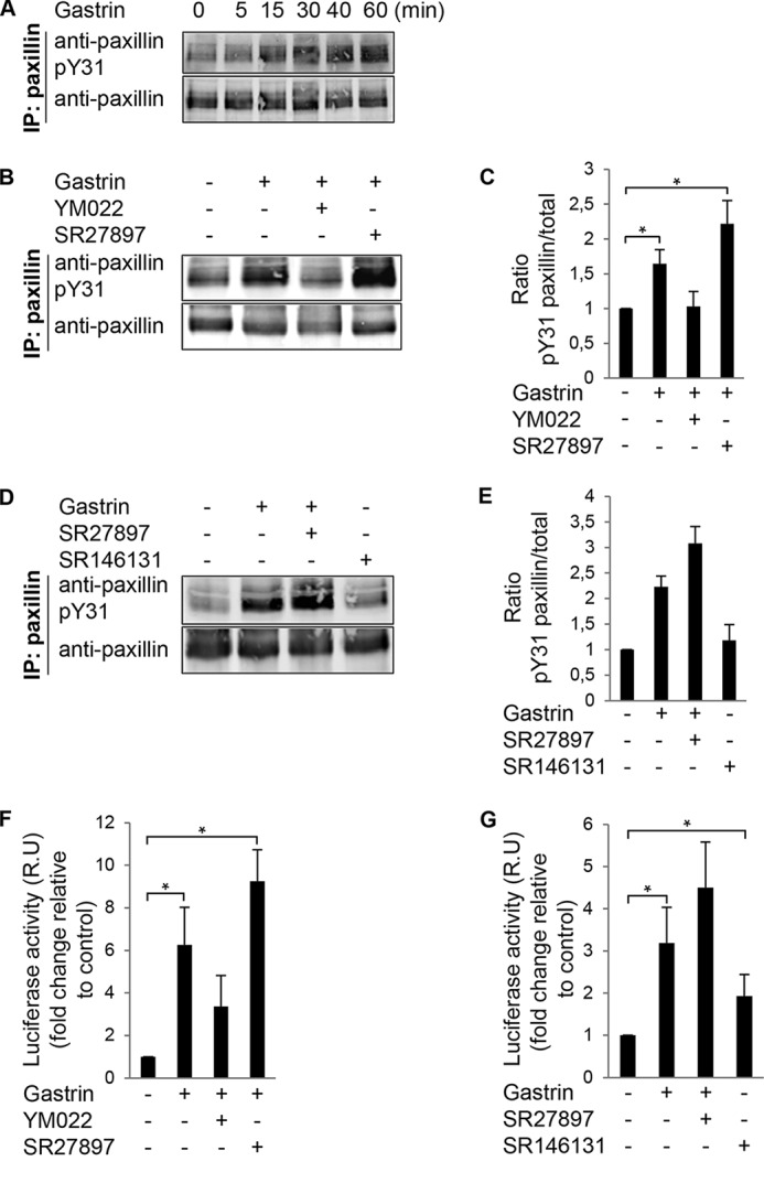
Gastrin stimulation of paxillin tyrosine phosphorylation and SRE activation in DLD-1 cells. A, gastrin (200 nm) was added to DLD-1 cells for the indicated times. Protein lysates were analyzed by paxillin IP and phosphospecific paxillin (pY31) or total paxillin immunoblotting. B, DLD-1 cells were pretreated with 2 μm SR27987 (CCK1R antagonist) or 2 μm YM022 (CCK2R antagonist) for 20 min. Gastrin (200 nm) was added, and lysates were prepared for paxillin IP after 40 min as in A. C, LI-COR image quantification of Tyr(P)-31 paxillin to total paxillin ratio from experiments in B. Values were set to 1 and are means ± S.E. fold increase of three experiments conducted in triplicate (*, p < 0.05, two-tailed t test). D, DLD-1 cells were pretreated with 2 μm SR27987 (CCK1R antagonist). Cells were pretreated with gastrin (200 nm) or SR146131 (CCK1R agonist) for 40 min before anti-paxillin IP as in A. E, image quantification of Tyr(P)-31 paxillin to total paxillin ratio from experiments in D. Values were set to 1. Data are the mean ± S.E. fold increase for three independent experiments. F, DLD-1 cells were transfected with pSRE.L and Renilla luciferase (RLuc) vectors. Cells were serum-starved and then pretreated with 2 μm SR27987 (CCK1R antagonist) or 2 μm YM022 (CCK2R antagonist) for 20 min. Gastrin (200 nm) or vehicle was added for 5 h, and lysates were prepared for luciferase detection. G, DLD-1 cells were transfected as above and were pretreated with 2 μm SR27987 (CCK1R antagonist). Gastrin (200 nm) or SR146131 (CCK1R agonist) was added as indicated for 5 h, and lysates were prepared for luciferase detection. F and G, values (mean ± S.E. of three independent experiments in triplicate) were normalized based on expression of RLuc and expressed (relative units, R.U.) as fold induction over serum-starved conditions (*, p < 0.05, two-tailed t test).