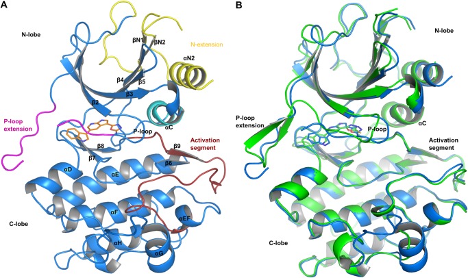
FIGURE 2.
Overview of the COT kinase structures. A, the overall structure of the COT·compound 2 structure is shown as graphic representation (Cα trace of the protein, blue). Compound 2 (orange, stick representation) binds to the active site of the COT kinase domain. N-extension, αC-helix, P-loop insert, and the activation segment are highlighted in yellow, cyan, magenta, and red, respectively. B, structure alignment of the COT·compound 2 (blue) with the COT·compound 3 (green) crystal structure. The Cα atoms of both structures were superimposed to generate this alignment (root mean square deviation = 0.454 Å2) Considerable conformational differences occur in the N-lobe of the COT kinase domain, in particular in the P-loop end P-loop insert segments.

