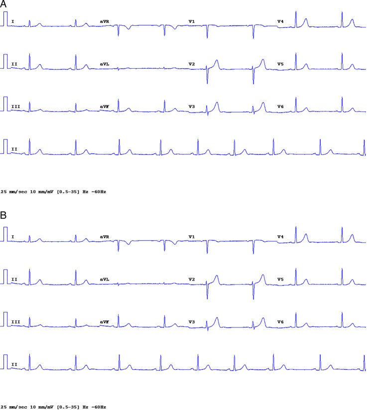
Figure 5.
(A) Standard 12-lead ECG: low R wave amplitude in lead 1, aVL; a test case chosen to flaw the new electrode placement (see online supplementary file, appendix 2, eFigures 1 to 32 and appendix 3 eFigure 21, 22). (B) New electrode placement ECG; similar to that of the standard ECG. The R wave amplitude in all 12 leads is virtually the same. The very small R wave in aVL did not change to a QS pattern (see online supplementary file eAppendix 2, eFigures 1 to 32).

