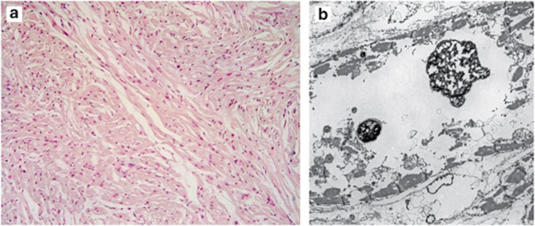
Figure 1.
(a) Microscopic postmortem examination of heart muscle from patient 1. Hypertrophic myocytes with myofibrillar disarray typical of HCM due to sarcomeric protein variations were present, albeit without a significant amount of interstitial fibrosis (hematoxylin and eosin staining, magnification × 100). (b) Electron micrograph showing a large, irregular vacuole.

