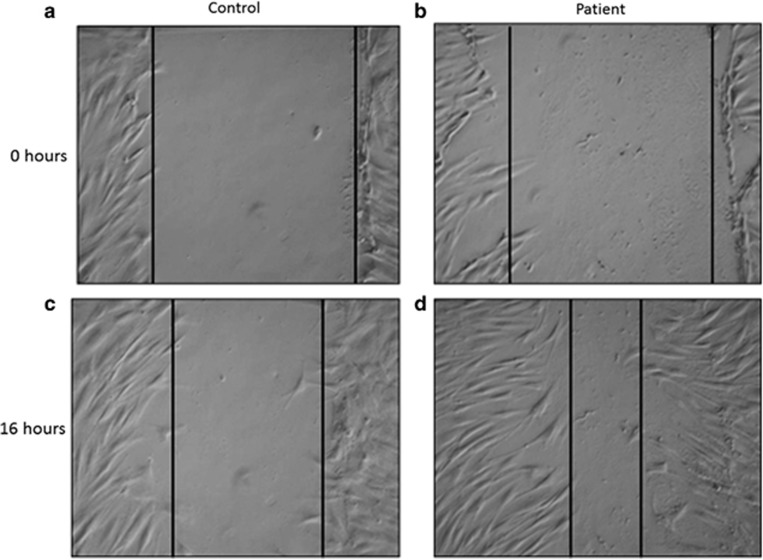
Figure 2.
Wound-healing assay performed on control and patient's fibroblasts: control fibroblasts (a) and patient's fibroblasts (b) immediately after the wound; control fibroblast (c) and patient's fibroblasts (d) 16 h after the wound. Vertical lines delimit the wound, with an equal width in (1, 2), whereas patient's fibroblasts exhibit a greater ability to migrate than do control cells, 16 h after the wound (c, d).

