
Figure 27.
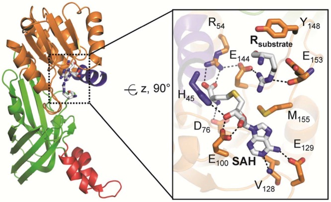
Active site architecture of PRMT1 bound to arginine (PDB code 1OR8). The structural elements are color coded according to Figure 25. The image on the right depicts details of the PRMT1 active site, highlighting critical residues for substrate binding and catalysis. Polar contacts of <3.5 Å are represented as dashed lines.