
Figure 30.
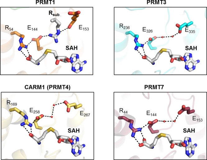
Structural representation of the active site of rat PRMT1 (PDB code 1OR8), rat PRMT3 (PDB code 1F3L), rat PRMT4 (PDB code 3B3F), and mouse PRMT7 (PDB code 4C4A). All structures contain a bound cofactor (SAH, highlighted in gray). Abbreviation: Rsub, substrate arginine.