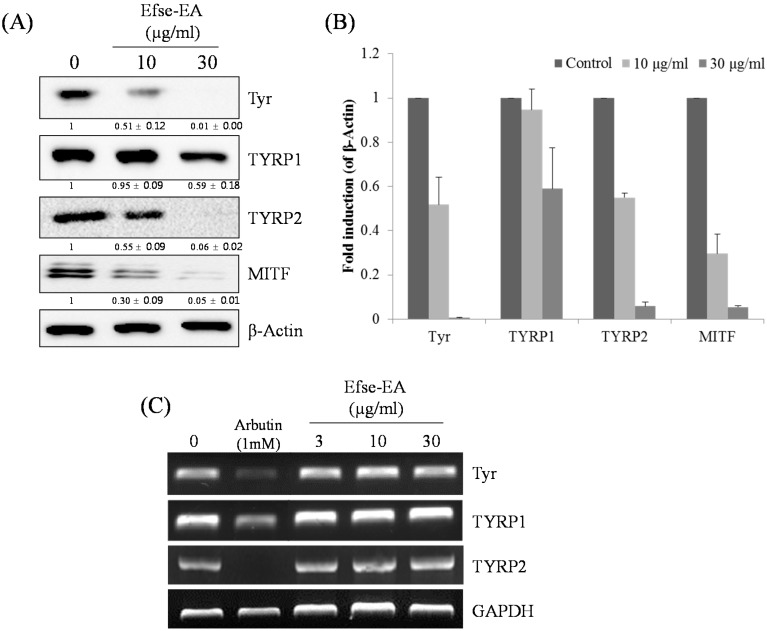
Figure 5.
Effect of Efse-EA on the levels of melanogenesis-related mRNA and proteins in melan-a cells. Cells (1 × 10⁵ cells/mL) were cultured for 24 h; the medium was replaced with fresh medium containing the indicated concentrations of Efse-EA or arbutin for three days. Total cell lysates were extracted and assayed by Western blotting using antibodies against tyrosinase, tyrosinase-related protein (TYRP)-1, TYRP-2, and microphthalmia-associated transcription factor (MITF). Equal amounts of protein loading were confirmed using β-actin (A); Relative intensity of melanogenesis-related protein expressions, the intensity of the protein expressions was compared to the control; The normalized data for each were plotted as bar graphs (B); Cells (1 × 10⁵ cells/mL) were cultured for 24 h; the medium was replaced with fresh medium containing the indicated concentrations of Efse-EA or arbutin for 1 day. The mRNA was extracted using TRIzol; mRNA expression was analyzed by reverse-transcription polymerase chain reaction (C).

