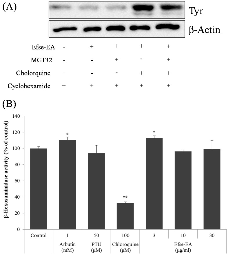
Figure 6.
Effect of Efse-EA on lysosomal tyrosinase degradation in melan-a cells. Cells (3 × 10⁵ cells/mL) were pretreated with 25 μg/mL cycloheximide for 1 h, as indicated. Cells were also pretreated with 10 μM MG132 or 50 μM chloroquine for 1 h, and then treated with Efse-EA for 6 h. Whole cell lysates were subjected to western blotting using an anti-tyrosinase antibody. Equal protein loading was confirmed using actin (A); β-Hexosaminidase assay was calculated as described in Experimental Section. In brief, melan-a cells (2 × 10⁵ cells/well) were seeded and sensitized with 1 μg/mL of dinitrophenyl (DNP)-immunoglobulin E (IgE) and stimulated with 20 μg DNP-bovine serum albumin (BSA). Following 1 h incubation, supernatant was transferred and the substrate for β-hexosaminidase (1 mM 4-nitrophenyl N-acetyl-β-d-glucosaminide (NAG; N9376, Sigma) was added. After adding stop solution, the sample was measured at 405 nm with a spectrophotometer (B). * p < 0.05, ** p < 0.01.

