Figure 1.
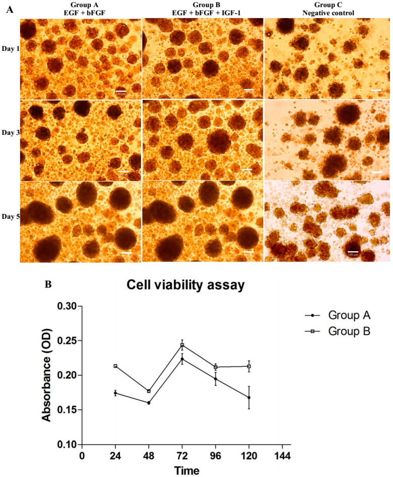
(A) Differentiation of bone marrow derived mesenchymal stem cells (BMSCs) into neural progenitor-like cells (NPCs) at day 1, 3 and 5 showing differing in sizes and morphology of neurospheres under different combination of growth factors. Cells were cultured in NeuroCult® NS-A neural basal media under serum free condition. Group A (EGF + bFGF), a published protocol of combination of growth factors required for neuronal differentiation of BMSCs; Group B (EGF + bFGF + IGF-1), an enhanced protocol of neuronal differentiation; and Group C (neural basal media without growth factor,) served as negative control of the experiment. Neurospheres with irregular shape was observed only in group C. Images were viewed under inverted light microscope with 100× magnification, Scale bar = 150 µm; (B) Cells proliferation were studied at five time intervals (24, 48, 72, 96 and 120 h) with 4 h incubation each with CellTiter 96® Aqueous One solution reagent (n = 6). Group B contained significant higher proportions of vial cells (p = 0.0098 with 95% CI) compared to Group A. The data represented as optical density (OD) at 540 nm in mean ± standard deviation.

