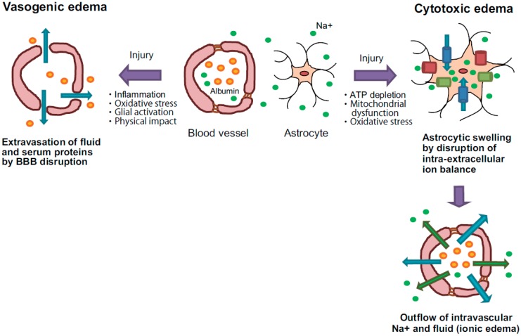
Figure 1.
Pathology of vasogenic and cytotoxic edema. Vasogenic edema: After brain injuries, endothelial tight junctions are disrupted by inflammatory reactions and oxidative stress. Moreover, activated glial cells release vascular permeability factors and inflammatory factors, and these factors accelerate blood-brain barrier (BBB) hyperpermeability. These events cause extravasation of fluid and albumin, leading to extracellular accumulation of fluid into the cerebral parenchyma. Cytotoxic edema: Brain insults induce intracellular ATP depletion, resulting in mitochondrial dysfunction and oxidative stress. These events cause a disturbance of intra-extracellular ion balance. As a result, excessive inflows of extracellular fluid and Na+ into cells are induced, leading to cell swelling. Because the extracellular Na+ contents are decreased by excessive inflow into cells, the outflow of Na+ and fluid from blood vessels is compensatorily accelerated. The intravascular Na+ outflow results in extracellular fluid accumulation in the cerebral parenchyma. Blue arrows: flow of water, green arrows: flow of Na+, orange spheres: albumin, green spheres: Na+, blue columns: water channel, green columns: ion transporter and red columns: ion channel.

