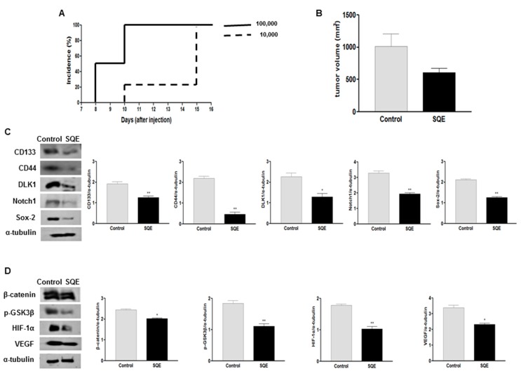
Figure 8.
Effect of SQE on tumorigenicity and CSC marker expression of CD133+CD44+ HT29 cells in vivo. (A) The tumorigenicity of CD133+CD44+ double-sorted cells (10,000 or 100,000 cells) was examined following the subcutaneous injection of these cells into nude mice; (B) Tumor volume was measured and compared between the control group and the SQE (300 mg/kg body weight) supplemented group; (C,D) Expression levels of various CSC markers, including CD133, CD44, DLK1, Notch1, and Sox-2 (C), as well as self-renewal and metastasis signaling-related markers, including β-catenin, p-GSK3β, HIF-1α, and VEGF (D), were detected by Western blot analysis. Detection of α-tubulin was used as a loading control. Representative figures are shown (left panel). Quantification of each protein level was shown with a bar graph. Bar represented the mean ± SEM. * and ** show significantly difference between two groups by Student’s t-test (p < 0.05). SQE, Sasa quelpaertensis leaf extract.

