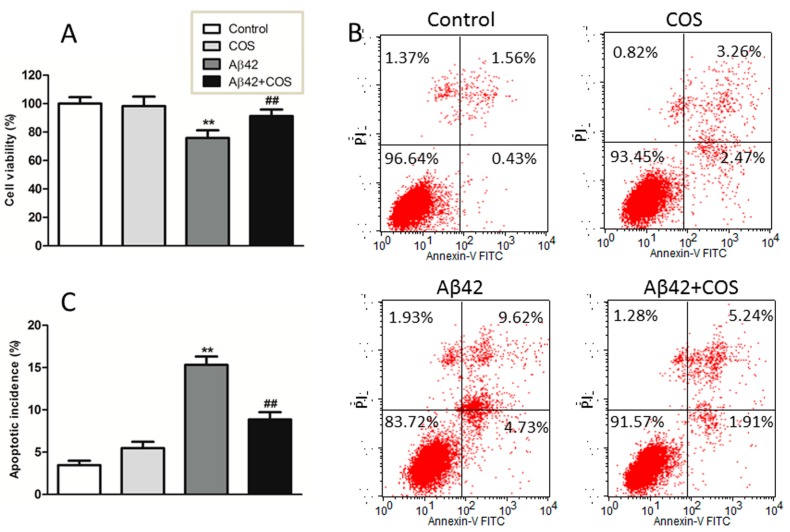
Figure 4.
Effect of COS on Aβ42-induced neurotoxicity in cortical neurons. (A) Neurons were treated with 5 μM Aβ42 with or without addition of 0.5 mg/mL COS for 48 h, the cell viability was determined by the MTT assay; (B) Representative graphs obtained by flow cytometry using double staining with Annexin V-FITC and PI; (C) The apoptotic incidence of rat cortical neurons exposed to 5 μM Aβ42 in the presence or absence of 0.5 mg/mL COS for 48 h. Results were expressed as the percentage of apoptotic cells that include neurons in early and late apoptotic phases. Data were expressed as means ± SEM of three independent experiments. ** p < 0.01 vs. Control, ## p < 0.01 vs. Aβ42 group.

