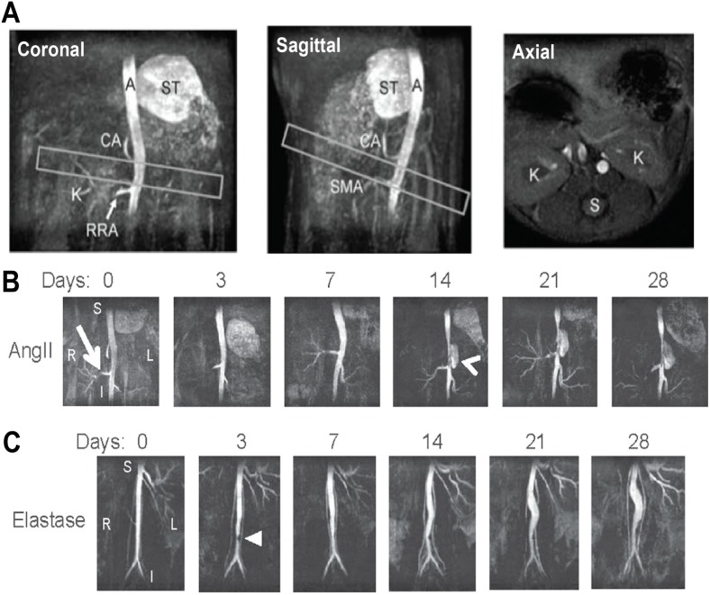
Figure 3.
Time-of-flight Magnetic Resonance Angiography (TOF-MRA) of Abdominal Mouse Aorta with and without Aneurysm. (A) TOF-MRA images of a healthy mouse aorta are shown as maximum intensity projections. Leftward position and antero-posterior curvature above the kidneys can be seen (boxes). The aorta (A), celiac artery (CA), kidney vasculature (K), right renal artery (RRA), spine (S), stomach (ST), and superior mesenteric artery (SMA) are labeled for landmark identification; (B,C) Coronal TOF-MRA maximum intensity projections show lumen expansion in (B) AngII and (C) elastase AAAs over 28 days. The AngII aneurysm appears suddenly (arrowhead) above the right renal artery (arrow) and expands leftward. The elastase aneurysm expands slowly, and a small region of signal hypointensity is observed due to a suture in the vessel at day 3 (triangle). Subfigure A is adapted with permission from Goergen et al. [108]. Subfigures B and C are adapted with permission from Goergen et al. [50].

