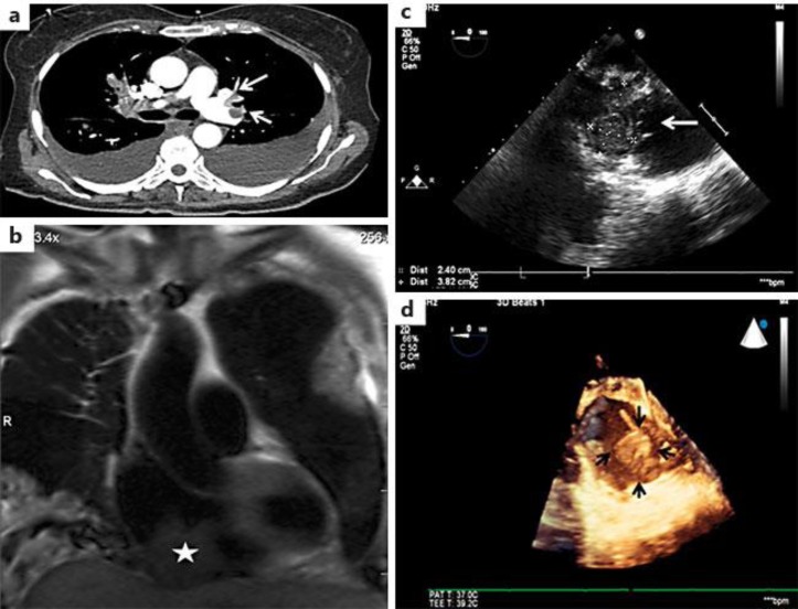
Fig. 1.
The patient had intracardiac metastases of HCC with a massive PE. a The CTA showed a bilateral effusion and extensive pulmonary embolisms involving the major branches of the left upper and lower lobes. b An MRI showed a large mass contiguous with the right ventricular and atrial septum. c The intraoperative transesophagal echocardiogram. d 3-dimensional echocardiography showed a large mass (about 6 cm) attached to the back wall of the posterior right atrium.

