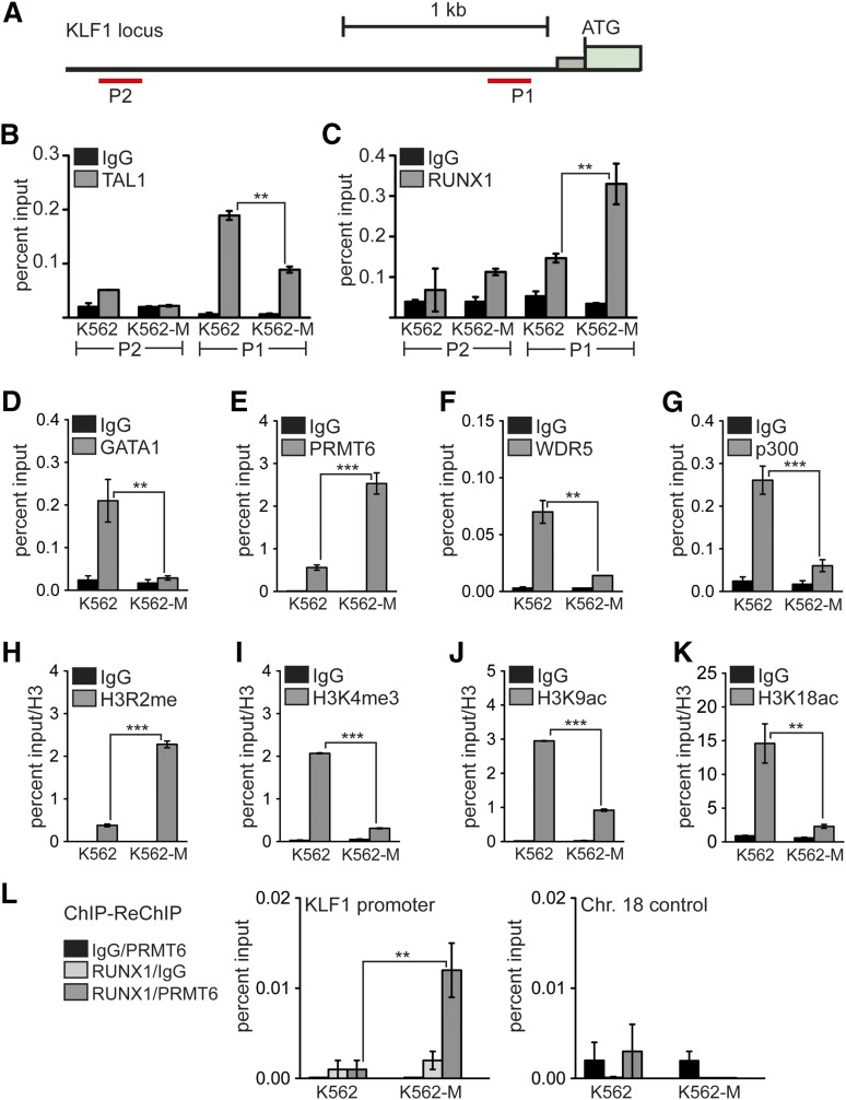
Figure 4.
Increased RUNX1 binding to the KLF1 promoter correlates with repression. (A) Schematic representation of the KLF1 promoter region. The position of primer pairs for ChIP is shown (P1 and P2). Transcription factors and histone modifications at the KLF1 promoter were measured in wild-type K562 cells (K562) and after megakaryocytic differentiation (K562-M) by ChIP. (B) Binding of TAL1 was detected at the P1 KLF1 promoter region in wild-type K562 cells (K562). TAL1 binding was reduced upon megakaryocytic differentiation (K562-M). (C) Increased RUNX1 binding was detected at the P1 region after megakaryocytic differentiation (K562-M). (D) GATA1 binding to the KLF1 promoter was decreased upon megakaryocytic differentiation. (E) PRMT6 binding to the KLF1 promoter was increased after megakaryocytic differentiation. (F) WDR5 binding to the KLF1 promoter was decreased upon megakaryocytic differentiation. (G) p300 binding to the KLF1 promoter was decreased upon megakaryocytic differentiation. (H) The H3R2me histone modification mark was increased after megakaryocytic differentiation. (I) H3K4me3 was reduced upon megakaryocytic differentiation. (J) H3K9ac was reduced after megakaryocytic differentiation. (K) H3K18ac was reduced upon megakaryocytic differentiation. (L) Quantitative ChIP-ReChIP of RUNX1 and PRMT6 with the given antibody combinations show co-occupancy of RUNX1 with PRMT6 at the KLF1 promoter (left) but not at a control region (right). Quantitative PCR values are shown as percentage input. Values gathered for histone H3 modifications were normalized with a ChIP against unmodified histone H3. The P values were calculated using Student t test. **P < .01; ***P < .001. ATG, start codon; Chr., chromosome.

