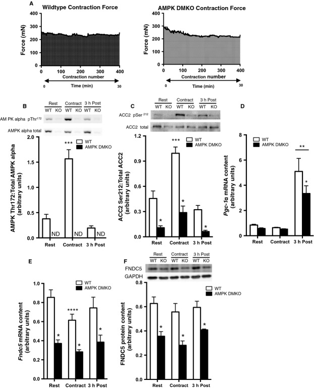
Figure 2.
FNDC5 expression in muscle from wild-type and AMPK DMKO mice at rest and after force-matched in situ contraction. (A) Tibialis anterior force generation over 400 contractions conducted over 30 min in wild-type (left) and AMPK DMKO mice (right). AMPK alpha pThr172 (B) and ACC2 pSer212 (C) phosphorylation levels, Pgc1α (D) and Fndc5 mRNA (E) and FNDC5 protein (F) content at Rest, immediately following (Contract), and 3 h post contraction (3 h Post) in the tibialis anterior muscle of wild-type and AMPK DMKO mice (N = 3–5). Error bars represent standard error of the mean. ND, not detectable. *Significantly different from wild-type, P < 0.05. **Significantly different from Rest and Contract, P < 0.05. ***Significantly different from Rest and 3 h Post, P < 0.05. ****Significantly different from wild-type Rest, P < 0.05.

