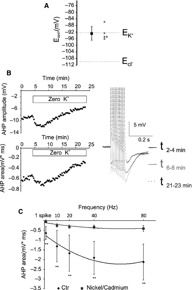
Figure 3.

The residual component of the AHP is mediated by K+ currents. (A) The reverse potential of the residual AHP is close to the calculated Ek. Empty circles represent the EAHP of single neurons, whereas the black squares represent the average value of EAHP ± SEM. (B) Removal of the extracellular K+ produced a transient increase, followed by a reduction, in the residual AHP. This specific pattern is possibly caused by the consecutive transient increase and sustained decrease in the potassium driving force due to the processes of extracellular and then intracellular potassium depletion. (C) The residual component of the AHP is reduced by the Ca2+ channel antagonists Ni2+/Cd2+. *Significant difference between the two conditions for each frequency. **P < 0.01.
