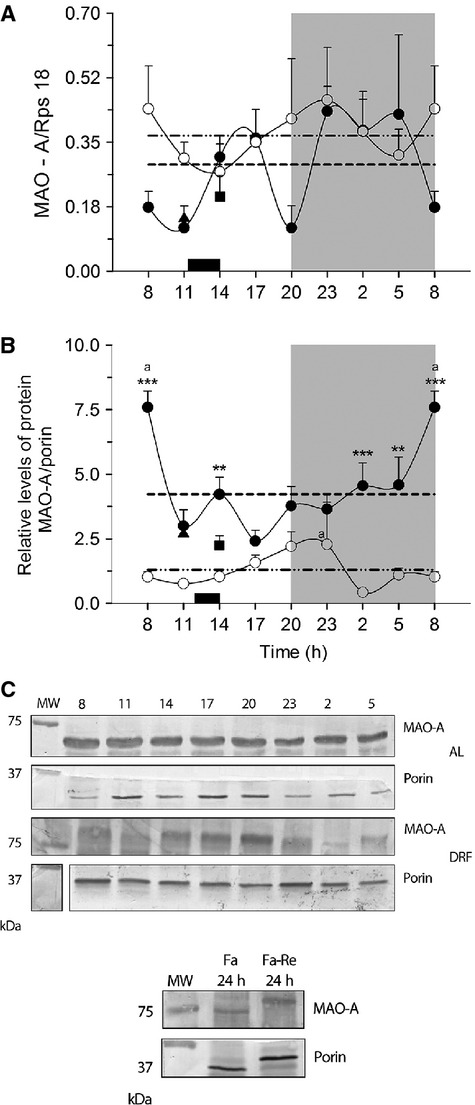
Figure 3.

MAO-A in the rat liver. (A) Levels of MAO-A mRNA expression and (B) protein in mitochondria (C) Blot representative sample of representative abundance of MAO-A protein in the different conditions. Each point represents the mean ± SEM. Access to food for the DRF group is represented by the black bar (1200–1400 h) AL (●), DRF (○), Fa (▲), Fa-Re (■), mesor AL(─) DRF (−∙∙), (a) Kruskall–Wallis test. *P < 0.05, **P < 0.01, ***P < 0.001, Kolgomorov–Smirnov two-samples test. (b) Mann–Whitney P < 0.05. n = 4. MAO, monoamine oxidase; DRF, daytime restricted feeding.
